A picture on the evolution of Serratia marcescens

Summary of: Sterzi, L., Nodari, R., Di Marco, F. et al. Genetic barriers more than environmental associations explain Serratia marcescens population structure. Commun Biol 7, 468 (2024). https://doi.org/10.1038/s42003-024-06069-w

In bacterial species we often observe a remarkable genetic diversity, with the presence of clear distinct genetic clusters. The view on how these genetic clusters are formed and maintained (i.e. speciation models) initially focused mostly on the interaction between mutations, selection and ecological divergence. These forces create a dynamic equilibrium where new lineages continuously emerge by mutation and compete for the same ecological niche, until one eventually out-competes the others. In this view, diversity emerges mainly when a mutation allows it to occupy a novel ecological niche: each lineage represents an “ecotype”. In the last decades, the dynamic nature of the bacterial gene repertoires and the extent of recombination (i.e. gene flow and horizontal gene transfer) in bacterial evolution have led to suggest a speciation model where genetic diversity is enhanced and maintained by barriers to recombination between subpopulations. In this study, we investigated how ecological divergence and genetic barriers could explain the population structure of Serratia marcescens, an opportunistic pathogen with high ecological and genetic plasticity. 

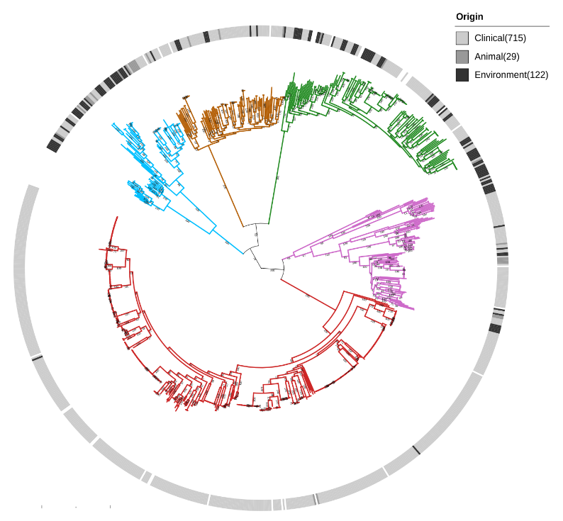
We show that Serratia marcescens comprises five deeply-demarcated genetic clusters. Analysing gene content and isolation sources, we observed that only two clusters show consistent signals of ecological specialisation, with one cluster strongly associated with clinical settings. We also used cluster-specific genes to investigate the occurrence of the genetic clusters in bacterial communities sampled from different biomes. We noted that the clusters co-occur in several biomes, but we also highlight that the clinically-associated cluster is enriched in freshwater samples, suggesting a possible reservoir. 

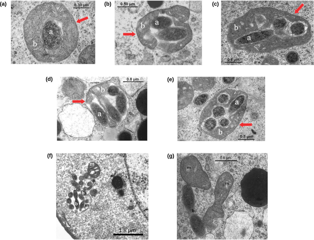
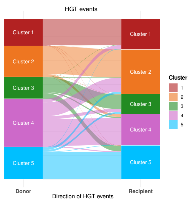
We studied gene flow within the species and, interestingly, we observed that genetic exchange between the five clusters in Serratia marcescens is notably limited. For this reason, we focused our attention on Restriction-Modification (R-M) systems. These systems are widespread bacterial defence systems which remove exogenous DNA, relying on a methyltransferase which methylates a specific sequence motif on the endogenous DNA and on a cognate restriction endonuclease which cleaves DNA when the motif is unmethylated. We observed a partial incompatibility of R-M systems coherent with limited inter-cluster gene flow, and we also noted that clusters are enriched in different types of R-M systems.

In conclusion, we believe that genetic barriers play a large part in maintaining diversity in subpopulation of S. marcescens. Within this population structure, two clusters have initiated adaptive trajectories to specific ecological niches and proceed to progressively isolate from the others. Whereas, other clusters are ecologically generalist and despite they often co-occur in the same environment at the same time, genetic barriers are sufficiently thick to maintain the clusters regardless of ecology or spatial distribution. Thus, with a hint of speculation, we propose that the leading role in the evolution of S. marcescens is played by the genetic barriers between co-occurring, ecologically generalist subpopulations.

The life cycle of Midichloria mitocondrii, a so interesting still open biological question

Summary of Comandatore F, … “Candidatus Midichloria mitochondrii” Using Electron Microscopy Data. mBio. 2021 Jun 29, DOI: 10.1128/mBio.00574-21.

During my master thesis period I worked on the genome assembly, annotation and phylogenomics of Midichloria mitochondrii, the first described intra-mitochondrial bacterium. M. mitochondrii is probably one of the most intriguing bacteria I ever met: it lives within the mitochondria of the oocytes of the hard tick Ixodes ricinus, occupying the space between the two membranes of the organelle. Surprisingly, the analysis of the M. mitochondrii genome led us to understand something important about the origin of mitochondria more than the evolution of the bacterium itself, but this is another story (see Sassera et al. 2011, https://doi.org/10.1093/molbev/msr159).

Years after I got the master degree, we had the opportunity to investigate one of the most interesting aspects of M. mitochondrii (in my opinion): its life cycle. Electron microscopy images clearly show mitochondrial cells colonized by one, two, three..up to about ten M. mitochondrii cells, and the highly “parasitized” ones having degraded structure. For this reason, it has been supposed that M. mitochondrii could behave similarly to a predatory bacterium. Following this hypothesis, the M. mitochondrii cells free in the oocyte cytoplasm invade the mitochondria and replicate within them until they lead to the organelle lysis (Sacchi et al. 2004, https://doi.org/10.1016/j.tice.2003.08.004).

To test this predatory-like hypothesis, we studied 71 I. ricinus oocytes from 11 ticks, counting the number of mitochondria parasitised by zero, one, two, three, … M. mitochondrii cells. Then, we used these values to model the bacterium life cycle within the oocyte. A total of 12,068 mitochondria and 7,805 “Ca. Midichloria mitochondrii” (intra-mitochondrial and free in the oocyte) units were observed.

Comandatore et al. 2021, https://doi.org/10.1128/mbio.00574-21

Surprisingly, the obtained results are not coherent with a predatory-like life cycle. We very rarely observed bacterial cells in replication, either within the mitochondria (two possible events observed) and in the cytoplasm (nine possible events). Furthermore, we observed a linear correlation between the frequency of cytoplasmic M. mitochondrii cells (normalized on the total number of oocyte M. mitochondrii) and the frequency of non colonized mitochondria (normalizes on the total number of mitochondria). In other words, we found that the more are the preys (i.e. non colonized mitochondria) the more are the predators (cytoplasmic M. mitochondrii). This peculiar linear relationship is quite unlikely in a pray-predators system, as we tested using the Lotka-Volterra model. Last but not least, it is very rare that a stable prey-predators system can emerge in a closed and limited space like an oocyte.

Thus, we try to change the point of view and we proposed another possible life cycle. Mitochondria have been found to be connected in a network in several organisms, in particular in humans. We hypothesized that the mitochondria of I. ricinus oocytes could be connected in a network and that M. mitochondrii could move, somehow, through it. Without experimental evidence we tested this hypothesis in silico using stochastic simulations. Our results show the the movement of
M. mitochondrii cells within an hypothetical mitochondrial network in coherent with the observed data: at the equilibrium, the number of mitochondria parasitized by zero, one, two, etc. bacteria are very similar to those observed by electron microscopy. 

Comandatore et al. 2021, https://doi.org/10.1128/mbio.00574-21

Can we say that M. mitochondrii moves through the oocyte mitochondrial network? At the state of the art, we can not be sure of this, also considering that our knowledge about I. ricinus mitochondrial network is very very limited. On the other hand, this work allowed us to understand that the predatory-like cloud is not the most fitting for
M. mitochondrii, while the bacterium could move within the I. ricinus mitochondrial network … even if this network really exists!

At the end, we scratched the surface of the problem and the life cycle remains an intriguing open question about this so interesting and difficult to study bacterium!